L2S Talk by Dr Joana Grah (Robert Koch Institute): Light and Electron Microscopy Image Analysis with Applications in Public Health Research
21 August 2025, 10.00 h in PB-H 0103
The talk is part of the ZESS lecture series and hosted by the DFG research unit “Learning to Sense”(L2S).
In this talk, we present some of the work currently conducted in the Image Analysis research group at the Centre for Artificial Intelligence in Public Health Research (ZKI-PH), Robert Koch Institute.
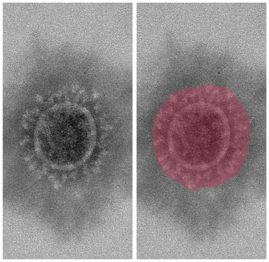
Microscopy imaging is an essential tool in biological and medical research. While light microscopy uses visible light to observe specimens such as living cells, tissues and microorganisms in real-time, electron microscopy achieves even higher resolutions using beams of electrons to visualise tiny objects such as viruses, enabling morphological analysis, detection of infectious agents and accurate diagnosis and understanding of disease mechanisms.
We show three use cases of data-driven imaging methods for this scenario: 1) CenSeFormer – a generalist model for cell segmentation in light microscopy, based on the novel CELLS data set, 2) Counting peplomers and measuring the viral envelope of SARS-CoV and SARS-CoV-2 viruses and 3) Automatic detection and classification of influenza A and borna viruses in cryo-electron microscopy images.
Dr Joana Grah is an applied mathematician and a keen science communicator based in Berlin. With a PhD in Applied Maths from the University of Cambridge, UK, her work has been focussing on the development of imaging tools for biomedical applications. Currently, she is working as a Research Associate and Deputy Leader in the Image Analysis group at the Centre for Artificial Intelligence in Public Health Research, which is part of the Robert Koch Institute.
During her visit at ZESS, the Women in Vision Initiative will also host Dr Grah for an informal lunch.
